
Types of skin spots are best read by colour first: brown spots are mostly melasma, sun spots or post-inflammatory pigmentation; the best-known white spots are vitiligo, tinea versicolor and pityriasis alba; and red spots include pityriasis rosea, eczema and psoriasis. Colour narrows the possibilities, but shape, texture, scale and location are what settle it, which is why some spots need an examination.
The Arabic pages that rank for this search list the types without pictures, and without explaining how to tell apart two spots of the same colour. This article is a map with pictures from published cases, the signs that separate the types, and when a spot needs a doctor.
Brown spots
Brown spots are the commonest. On the face, the best known are symmetrical melasma on the cheeks and forehead, well-defined sun spots, and freckles that darken in summer. On the body, add post-inflammatory pigmentation, acanthosis nigricans in skin folds, and the darker form of tinea versicolor. These are covered in detail in two articles: what causes brown spots on the face and brown spots on the body.
The one brown spot that is never treated cosmetically before it's examined is a changing mole: asymmetrical, with irregular edges, more than one colour, growing, or bleeding.
White spots: three common types
Tinea versicolor: a superficial fungus
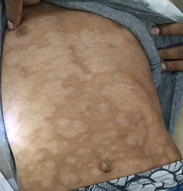
Tinea versicolor (pityriasis versicolor) is a superficial infection with a yeast that normally lives on the skin, more common with heat and sweating. It shows as lighter or slightly darker patches on the chest, back and abdomen, with fine scale that appears when you scratch gently, and it looks more obvious after tanning because the affected skin doesn't tan. The figure from a 2026 case shows extensive tinea versicolor on the abdomen:
It's treated with a topical or prescription oral antifungal, but normal colour returns slowly after the yeast has gone, and the infection can recur in summer. That slow return of colour is often mistaken for treatment failure; the fine scale disappearing is the better sign that it has worked.
Vitiligo: complete loss of pigment
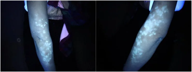
Vitiligo is a non-contagious autoimmune condition in which pigment cells stop working, leaving bright white patches with sharp edges and no scale, often symmetrical on the hands, around the mouth and eyes, and over joints. Under a Wood's lamp, an ultraviolet light used in clinic, vitiligo patches glow a distinct blue-white, as in this case from a 2025 report:
Vitiligo isn't contagious and isn't linked to food, but it can occur alongside other autoimmune conditions such as thyroid disease, so your dermatologist may order tests. Treatment options have widened in recent years, and starting early, while patches are still spreading, gives the best chance of repigmentation.
Pityriasis alba and sun-related white spots
Pityriasis alba is pale patches with blurred edges and fine scale, seen especially on children's faces after summer. It is linked to dry skin and eczema and fades on its own with moisturising. Very small white spots on the shins and forearms in adults are usually the result of long-term sun exposure and are harmless.
Red spots
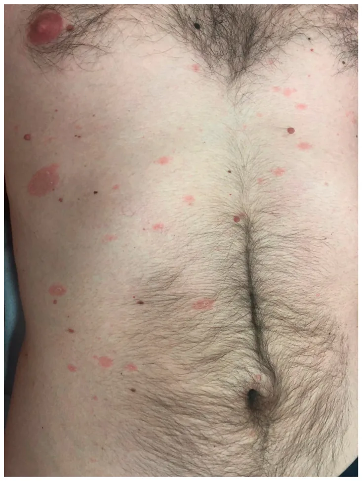
Red spots have the widest range of causes: eczema with itching and dryness, psoriasis with well-defined plaques and silvery scale, ringworm with a red ring and an active edge, and pityriasis rosea. Pityriasis rosea usually starts with one large pink patch on the trunk, followed days to weeks later by smaller patches, as in this figure from a 2025 review:
Pityriasis rosea usually clears on its own within weeks, but it can resemble ringworm or other conditions, and pityriasis rosea in pregnancy is worth discussing with your doctor. The most important red type is small red dots that don't fade when you press a clear glass against them, because they can mean bleeding under the skin and need urgent assessment.
A quick comparison
| Colour | Commonest types | Distinguishing sign | Next step |
|---|---|---|---|
| Brown | Melasma, sun spots, post-inflammatory pigmentation | Symmetry, edges, history of inflammation | Sunscreen and identifying the type |
| Bright white | Vitiligo | Sharp edges, no scale, glows under Wood's lamp | Autoimmune assessment and prescription treatment |
| Pale with scale | Tinea versicolor | Fine scale on scratching, chest and back | Antifungal |
| Pale, blurred | Pityriasis alba | A child's face, dryness | Moisturising |
| Red and scaly | Pityriasis rosea, psoriasis, ringworm | Herald patch, silvery scale, or a ring | Examination before any cream |
| Red dots that don't fade | Bleeding under the skin | Don't disappear with pressure | Urgent assessment |
flowchart TD
  A[A new spot] --> B{What colour?}
  B -->|Brown| C{Changing, irregular or bleeding?}
  C -->|Yes| D[Prompt examination]
  C -->|No| E[Sunscreen and identify the type]
  B -->|White| F{Fine scale on it?}
  F -->|Yes| G[Probably tinea versicolor]
  F -->|No| H[Vitiligo or another type - assessment]
  B -->|Red| I{Dots that don't fade with pressure?}
  I -->|Yes| D
  I -->|No| J[Examination before using any cream]
mindmap
  root((Skin spots))
    Brown
      Melasma
      Sun spots
      Post-inflammatory
    White
      Vitiligo
      Tinea versicolor
      Pityriasis alba
    Red
      Pityriasis rosea
      Eczema and psoriasis
      Ringworm
Why "just try a cream" goes wrong
Many spots are treated at home with whatever cream is to hand, and the results can be misleading. A steroid cream on ringworm calms the redness for a while but lets the fungus spread and changes how it looks, making diagnosis harder later. An antifungal on vitiligo does nothing. A lightening cream on a changing mole delays the one examination that matters. That is why the safest first step for any new spot you can't identify is to look at it properly, not to treat it.
Frequently asked questions
What are the types of skin spots?
In practice, skin spots are grouped by colour. Brown ones include melasma, sun spots, freckles and post-inflammatory pigmentation; white ones include vitiligo, tinea versicolor and pityriasis alba; red ones include pityriasis rosea, eczema, psoriasis and vascular spots. Colour is where the thinking starts, and an examination settles it.
What's the difference between vitiligo and tinea versicolor?
Vitiligo is bright white patches that have lost their pigment completely, with sharp edges and no scale, which glow under a Wood's lamp. Tinea versicolor is a superficial fungus that causes lighter or slightly darker patches with fine scale that shows when lightly scratched, and it's treated with an antifungal.
Are white spots on the skin dangerous?
Most aren't: tinea versicolor is a treatable superficial fungus, pityriasis alba fades on its own in children, and vitiligo isn't contagious. But vitiligo can be associated with other autoimmune conditions such as thyroid disease, and some spots need examining to rule out rarer causes.
When are skin spots serious?
When a spot changes in size, shape or colour, has irregular edges, bleeds or won't heal, or when small red spots that don't fade under pressure appear together with fever or bruising. These need prompt examination.
When to see a clinician
See a dermatologist if bright white patches are spreading, if scaly patches haven't improved, or if you have a spot you can't identify before putting any cream on it. See one promptly if a mole or spot changes in shape, colour or size, or bleeds. Seek urgent care if small red or purple dots appear that don't fade under pressure, especially with fever, tiredness, bruising or bleeding gums.
Related articles
- What causes brown spots on the face
- Brown spots on the body: causes and how to tell them apart
- The fastest way to remove facial pigmentation
- Melasma treatment for the face
Further reading
- Tinea versicolor: Signs and symptoms — American Academy of Dermatology
- Vitiligo: FAQs — American Academy of Dermatology
- Pityriasis rosea: FAQs — American Academy of Dermatology
Medical disclaimer: This article is for health education only and is not a substitute for professional medical advice. If you have a skin condition that concerns you, please consult a dermatologist.